Flow cytometers are valuable in the laboratory because of their ability to rapidly analyze thousands of normal cells while being able to identify the presence of rare and abnormal cells. Combining flow cytometry testing with other laboratory tests and patient history is critical to a clinician’s ability to diagnose certain types of diseases, establish a prognosis, monitor response to treatment and maintain vigilance for relapse. Advancements in flow cytometry technology are leading to increases in the number and types of flow cytometry tests being routinely run in the clinical lab and flow cytometry will continue to grow in importance.
Flow cytometry testing is performed in both clinical and research labs. Common tests performed by flow cytometry include immunophenotyping, CD4+ T cell counts for HIV/AIDS monitoring, hematopoietic stem cell enumeration for stem cell transplants, HLA (human leukocyte antigen) typing for stem cell and organ transplants, leukemia/lymphoma classification and minimal residual disease (MRD).
Early days of flow cytometry
In the 1950s, the only laboratory tool available for the assessment and characterization of biological tissue at the cellular level was optical microscopy. Then a method using laminar flow and hydrodynamic focusing was developed to “see” red and white blood cells. These first “flow cytometers” were only capable of measuring cell size by impedance calculations based on the Coulter principle. When this flow-based technology was paired with fluorescently labeled antibodies and sensitive detectors in the 1970s, modern flow cytometry was born. This represented a major technological step forward and began a rapid development of this new technology that continues to this day and is likely to continue well into the future.
In the decades since its invention, flow cytometry has flourished to provide a wide set of capabilities that are unique to this powerful diagnostic procedure, far exceeding those of microscopy. Now, flow cytometry can identify and quantify up to 30 or more cell parameters at a time, while microscopy – even with special instrumentation – can only identify six.
What is flow cytometry?
Flow cytometry is a rapid method used to detect, identify and measure multiple characteristics of an individual cell. Flow cytometry can be used to evaluate cells from virtually any kind of biological sample, such as:
- Peripheral blood
- Cerebrospinal fluid (CSF)
- Bone marrow
- Biopsies from tumors and lymph nodes
- Urine
Flow cytometry is based on simple principles that, when combined, form a powerful analytical tool.
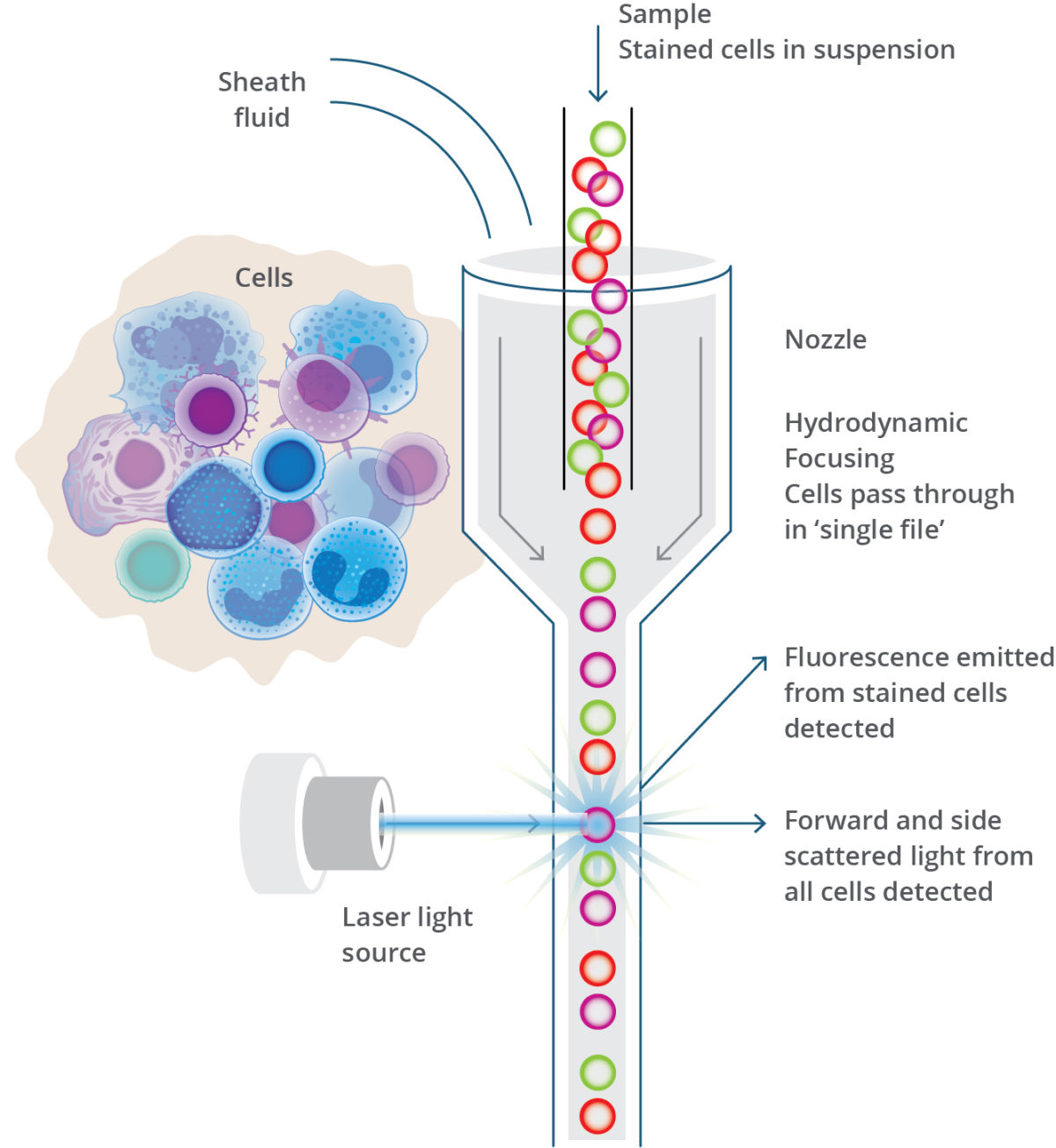
Cells are incubated with fluorescently labeled antibodies that bind to antigens of interest on the cell. The cells are then suspended in a neutral fluid and analyzed on the flow cytometer. The flow cytometer takes up the fluid and cells, and they are organized by laminar flow so that they pass through a chamber, called a flow cell, one cell at a time. As the cells pass through the flow cell, a laser beam excites the fluorophores each time a cell passes through its path; this is called an event. The light-scatter from the cell and emitted fluorescence from the antibody-fluorophore combination is then collected by optical detectors and is transferred to a digital format to be analyzed on a dot plot or histogram.
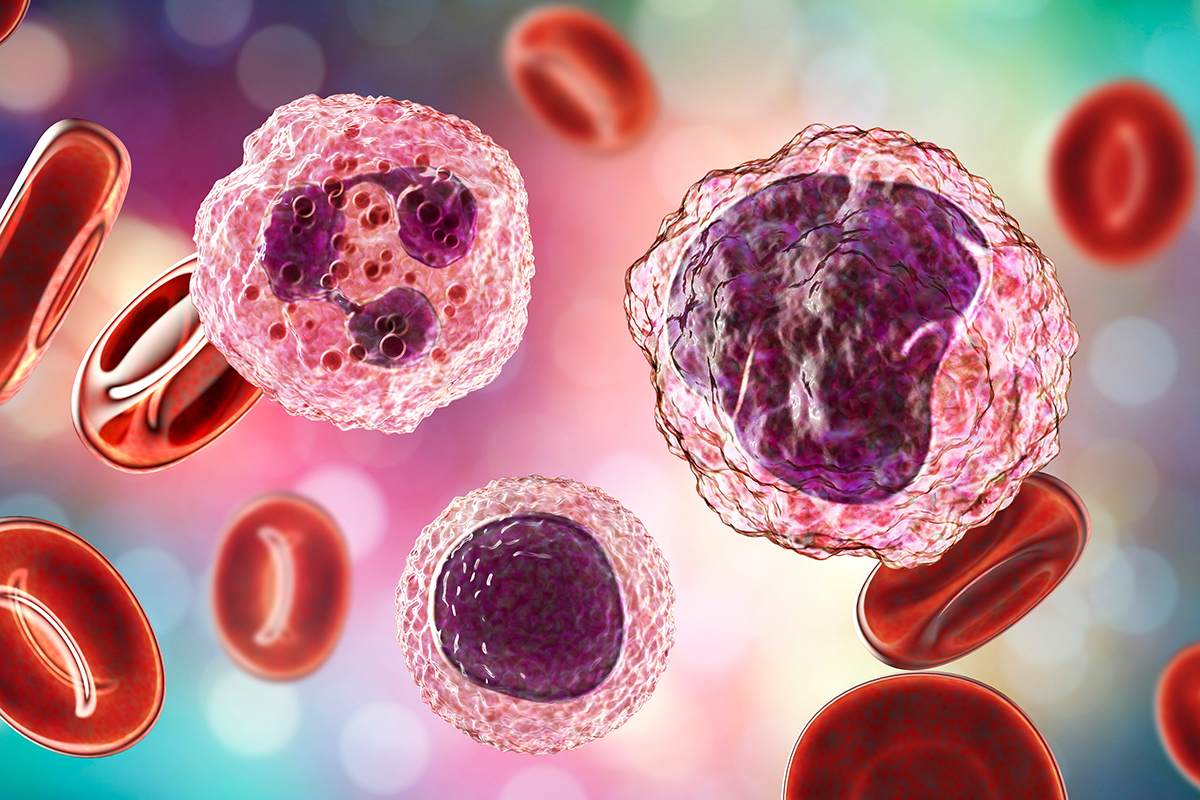
Light scatter collected by forward (FCS) and side scatter (SSC) creates patterns that are characteristic of the size and internal complexity of the cell. This indicates what type of cells (ex., lymphocyte, monocyte, granulocyte, platelet) are present.
The fluorescent labeled antibodies identify the cell’s surface and cytoplasmic antigens which are often designated as clusters of differentiation (CD) markers. These CD markers can also be used to associate cells with certain immune functions and health of the patient. Any aberrant expression of the CD markers leads clinicians to investigate the cause of the abnormality. Flow cytometry testing is performed in both research and clinical labs. The information obtained by “interrogating” cells can be extremely useful for purposes of diagnosing and monitoring disease.
Glossary of common flow cytometry terms

The clinical lab must maintain CAP accreditation and CLIA compliance by running positive procedural controls to verify the performance of reagents, preparation methods, staining procedures and instrument performance.
Streck offers the broadest portfolio of flow cytometry controls on the market. These controls not only provide values for both normal and abnormal leukocyte populations and reference values for intracellular and surface markers, but also have extended shelf life that will limit the number of lot-to-lot correlations and reduce the need to purchase controls from multiple companies. With many labs experiencing decreasing reimbursement for flow testing, you can maximize your lab’s efficiency with Streck flow controls.
Antibody ‒ a protein produced by the immune system in response to a foreign substance, often a virus or bacteria, called an antigen. The antibody recognizes and binds to this substance like a lock to a key and aids the immune system in targeting the foreign substance for destruction.
Antibody Titration ‒ the method of determining the correct amount of fluorophore conjugated antibodies to use in order to get the best resolution for each antigen and cell of interest.
Antigen ‒ generally a foreign substance, such as a chemical structure, bacterial protein, virus or pollen that can stimulate a specific immune response.
Autofluorescence ‒ non-specific fluorescence emission observed when certain cell native cell molecules are excited by a particular wavelength and give off light that is detected by the flow cytometer.
Clones ‒ genetically identical daughter cells from a unique parent cell.
Clusters of Differentiation (CD) ‒ a nomenclature system given to mostly surface proteins of the immune system in order to standardize naming and prevent confusion. Each unique surface molecule is assigned a different number which allows cell phenotypes to be identified. CD molecules may or may not be specific for just one cell or cell lineage. The official listing of determinants has identified more than 400 individual and unique markers, with more added every few years.
Density Plot ‒ a graphical representation of two-parameter data where the colors represent the number of events at that location. Each axis represents the single intensity of one parameter.
Dot Plot – graphical representation of two-parameter data where each axis represents the single intensity of one parameter and each dot represents a single cell’s level of that parameter.

Event ‒ an individual particle, generally a cell, that goes through the laminar flow cell and is interrogated by the flow cytometer for size, granularity and fluorescence levels.
Fluorophore/Fluorochrome ‒ a structure from a chemical or protein that accepts light (excitation) and re-emits it at a longer wavelength (emission), producing fluorescence. This is the basis of flow cytometry, which recognizes the wavelengths of interest. They are attached to monoclonal antibodies that bind to specific target cells or cell components.
Gates ‒ numerical or graphical boundaries that are drawn around cells with similar characteristics, such as forward light scatter (FCS), side light scatter (SSC) and marker expression (CD) to identify cells to further analyze.
Histogram ‒ a graphical representation of single-parameter data that shows relative number and distribution of events (or cells). The horizontal axis corresponds to the single intensity of a selected parameter, while vertical axis represents the number of events (counts).
Immunophenotyping ‒ the detection of specific cell types by using flow cytometry to identify immune-related markers on the cell surface. This is the basis of the most common clinical uses of flow cytometry.
Resolution ‒ the potential of a particular flow cytometry instrument to detect differences in fluorescence frequencies.
Maximize your lab’s efficiency with Streck flow controls