Clinical lab body fluid evaluation
Topics Featured
Contributing author: Jessica Hart, Streck RD Associate Scientist 2
To paint a clear diagnostic picture for many pathological conditions, healthcare providers often request the collection and clinical laboratory evaluation of more than just blood samples. The results from body fluid analysis can provide useful diagnostic clues for clinicians in cases such as inflammation, infection, injury, malignancy, autoimmune disorders and pressure imbalances.
Some common body fluids submitted to clinical laboratories for analysis include cerebrospinal fluid (CSF), synovial fluid, serous fluids (pleural, peritoneal or pericardial effusions) and respiratory fluids (bronchial alveolar lavages, or BALs). Evaluation of body fluids typically includes white blood cell (WBC) and red blood cell (RBC) counts, cell identification and/or differential counts, crystal identification, tests for microbial contamination and chemistry analysis.
Quality control practices in clinical laboratories should align with CLIA, CLS and CAP guidelines, meaning a quality control sample must be evaluated for cell counts at least once every 8 hours during which patient testing takes place. Cell counts for these quality controls must fall within ranges of expected values before patient cell counts can be released. However, body fluid evaluation typically includes more than just WBC and RBC cell counts on a hemacytometer.
Cells from body fluids may also be condensed into a small area of a slide (called a cytospin) and stained for microscopic visualization and evaluation of cellular morphology. This is done to enumerate relative increases/decreases in certain WBC types (seen in infections, inflammation or immune disorders) or to look for unusual, malignant cell types associated with cancer. Polarized light microscopy is also used to evaluate synovial (joint) fluids for presence of gout or pseudogout crystals.
A superior body fluid control
Good quality controls are designed to ensure the protocols and test methods used for patient samples consistently produce accurate and precise patient results. Patient lab results are an integral part of medical diagnoses, providing clinicians with valuable insight into the state of health for each patient. Quality controls are the first line of defense to ensure test systems are functioning optimally before patient results may be reported and have an impact on medical decisions. When patients and their families are relying on your lab, how will you be sure you’re providing them with high-quality results?
Streck Cell-Chex® body fluid control contains stabilized human cells preserved to resemble patient samples more closely than other available controls on the market, increasing confidence in the cell identification skills of laboratory technologists. Biconcave RBCs and morphologically distinct WBCs make cell identification easy and enable the recovery of accurate cell counts within expected assay ranges.
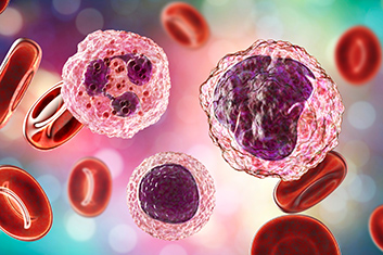
Cell-Chex is the only manual procedural control that checks more aspects of body fluid evaluation than just WBC and RBC counts. Its composition is representative of multiple body fluid types including CSF, serous fluids and synovial fluids (unlike other available controls which only claim to resemble CSF) and it has five morphologically distinct types of WBCs for performing differentials. When stained in the same manner as patient samples, neutrophil, monocyte, lymphocyte, eosinophil and basophil populations can be easily differentiated by laboratory professionals. Cell-Chex is also the only manual body fluid control that contains real urate and CPPD crystals for identification in synovial fluid samples via polarized light microscopy.
Cell-Chex cytospin preparation and staining protocols can be used to make slides exactly as patient slides are made, increasing confidence in slide preparation and staining techniques. In addition, microscope performance is checked during hemacytometer counts, WBC differentials and crystal identification (including polarized light capabilities), increasing confidence in the quality of microscopic analysis.
Click here for more information on Cell-Chex.

Jessica Hart is a certified Medical Laboratory Scientist and Specialist in Hematology, MLS(ASCP)SH, with a Master of Public Health in biostatistics. She previously worked in the clinical hematopathology laboratory at Nebraska Medicine before joining Streck in 2019. Her interests include hematology, flow cytometry, biostatistics, and all things related to the advancement of clinical laboratory science.


ILQC: How to earn the trust of your clients and their patients

